Medaka embryos

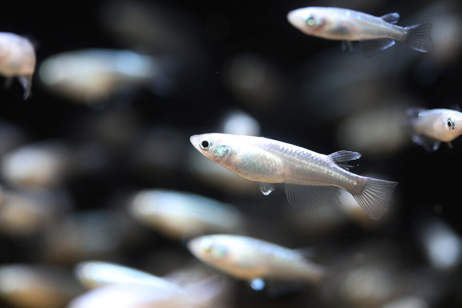
This project aims to develop a highly detailed description of the pituitary gland of the medaka fish. 

Our main objective

This project provides an overview of the mRNA content of each pituitary cell types in the male and female adult medaka.

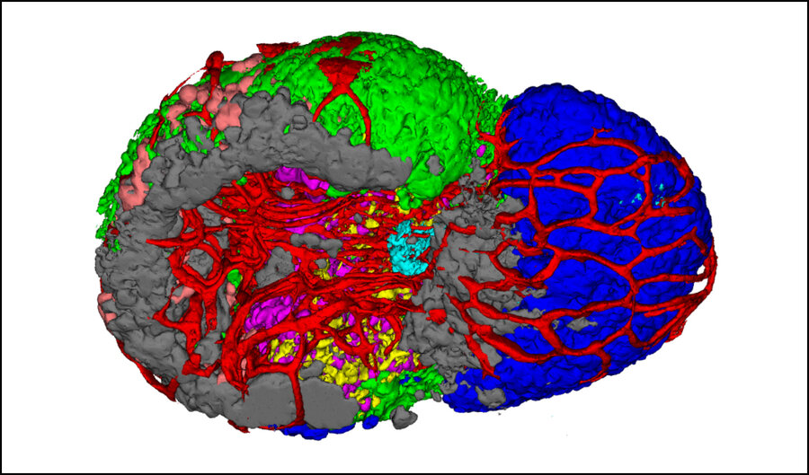
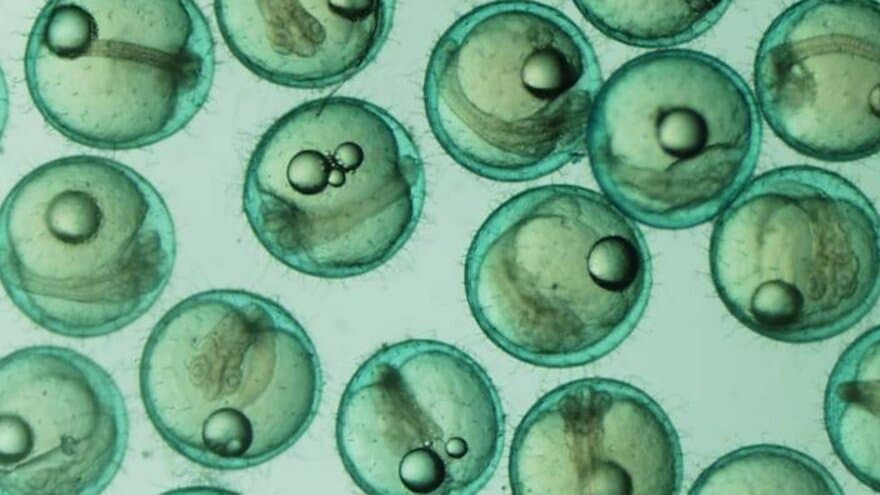
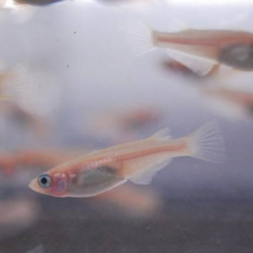
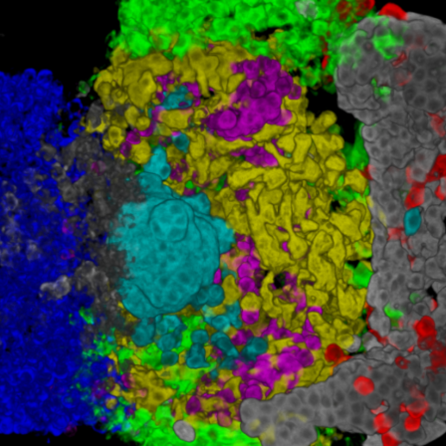
It also provides a detail 3D map of the distribution of the different endocrine cell populations in the juvenile and adult medaka.

  • This project aims to provide an overview of the location and transcriptomic identity of the different endocrine cell types in the medaka pituitary.

    The pituitary is a key endocrine grand in the central nervous system which participate in the regulation of essential physiological functions, such as growth, reproduction and stress response. Located below the hypothalamic region of the brain, the pituitary is divided into two main parts with different developmental origins: the neurohypophysis (posterior pituitary) and the adenohypophysis (anterior pituitary). The neurohypophysis, which come from an extension of the diencephalic region of the brain mainly contains projections from the brain. The adenohyphysis originates from an up-growth of the pharyngeal ectoderm and endoderm and can be histologically divided in the pars intermedia (PI), the pars distalis (PD), and the pars tuberalis (PT), the latter present in mammals but not in teleosts. The adenohypophysis contains several endocrine cell types. 

    Pituitary is a tissue that shows high plasticity, with the number of specific cell types changing through the life of an animal. The activity of the cells, hormone synthesis and release which are mainly regulated by neuroendocrine signals from the brain and by feedback regulatory signals from the target organs, is also regulated to adjust hormone production to the physiological needs. 

    The Japanese medaka:

    The Japanese Medaka (Oryzias latipes), is a small teleost fish similar to zebrafish in many aspects. However, medaka possess a smaller genome and a genetic sex determination system. He is also more tolerant in term of water salinity and temperature, which makes it a perfect model for many studies.

    Jan G. Bjaalie
  • This atlas provides a detailed 3D map of the distribution of different endocrine cell populations in the juvenile and adult male and female medaka pitutary.

    Maja Amedjkouh Puchades

    Description:

    Using fluorescent multi-color in situ hybridization technique, we developped 3D atlases of juvenile and adult male and female medaka pituitary. The atlas provides localization of seven endocrine cell types: lactotropes, thyrotropes, two distinct gonadotropes, somatotropes, somatolactotropes, and corticotropes + melanotropes. 

    This platform is user friendly, which enables the user to navigate to the whole pituitary from any angles and points of view as well as allows the user to focus on certain cell types of interest. Considering less detailed information provided by previous 2D maps of teleost pituitary, we introduce these 3D atlases 

  • Short video presentation explaining how to use the 3D model and how to navigate and open z-stack files in order to observe specific labels.

    1. Navigate with the 3D model

    2. Navigate to the repository of raw data files

    3. Make a 3D view of a raw data file

  • This single cell transcriptomic provides a detailgene expression profiles for 2592 and 3804 individual cells from the pituitaries of female and male adult medaka.

    Description:

    Using single cell transcriptomic approach from the 10X genomic platform we provide the  gene expression profiles for 2592 and 3804 individual cells from the pituitaries of female and male adult medaka (Oryzias latipes), respectively. Based on expression profile clustering, 15 and 16 distinct cell types were defined in the female and male pituitary, respectively, of which nine are involved in the exclusive production of a single peptide hormone. One cell cluster appears to be unique to the male pituitary. Collectively, our data provide a high-quality reference for studies on pituitary biology and the regulation of hormone production, both in fish and in vertebrates in general. 

    How to investigate the dataset:

    The main data record consists of barcodes.tsv, features.tsv and matrix.mtx files, listing raw UMI counts for each gene (feature) in each cell (barcode) in a sparse matrix format. In addition, we provide a table of cell type assignments and UMAP projections for each individual cell (barcode), as well as a summary of the top differentially expressed genes per cell type. Raw sequencing data are available in FASTQ format. Finally, the data record contains matrix files on exonic and intronic expression, which can be used for Velocyto gene expression dynamics analyses.

    We then invite you to follow the tutorial made by Satijalab to start with Seurat to use our R code available  on GitHub to open and process the data set.

Participants

Publications

2021

2020

News from the project from nmbu.no (in Norwegian)