The Imaging Centre
Forskningsinfrastruktur for avansert mikroskopi
En fullstendig tjeneste som inkluderer prøvepreparering, opplæring og bruk av mikroskop.
Veiledning og teknisk hjelp av hele vår infrastruktur, uavhengig av forkunnskaper.
Vi er tilgjengelige for alle interne og eksterne brukere, både fra akademia, industri og privatpersoner.
Imaging Centre har flyttet til Veterinærbygget, mai 2025.
Vi er tilgjengelige på e-post, så ta gjerne kontakt om det er noe.
Kontaktinfo:
E-post: imaging.centre@nmbu.no
Telefon: 67 23 27 61/62, 92 41 72 43 eller 48 25 98 43
Besøksadresse:
Elizabeth Stephansens vei 15, Hippocampus, Veterinærbygningen
Postadresse:
Postboks 5003 NMBU
1432 Ås
Se hvor du finner oss i MazeMap.
Åpningstider:
Kl. 08:00 til 15.45
15. mai til 14. september:
kl. 08:00 til 15.00
Nyheter
Booking og priser
Kurs
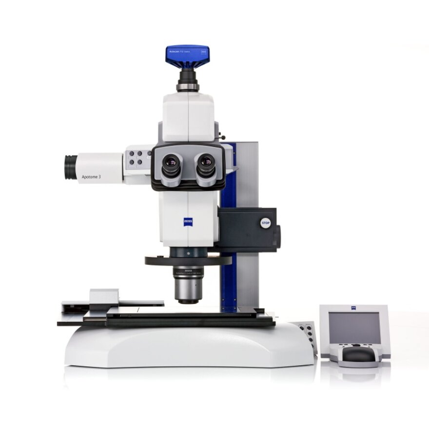
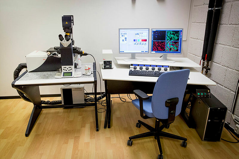
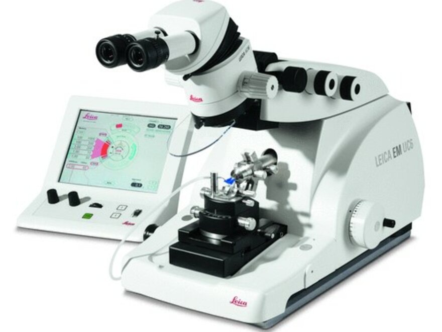
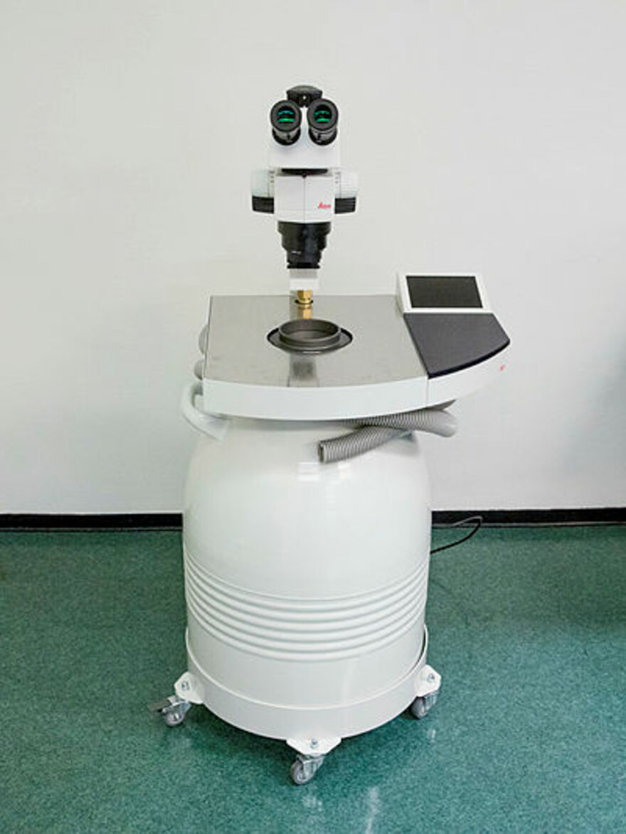
Utstyr


Ansatte ved Imaging Centre