The Imaging Centre
Core facility for advanced microscopy
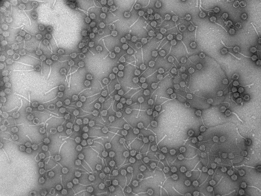
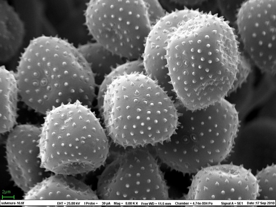
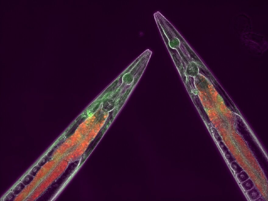
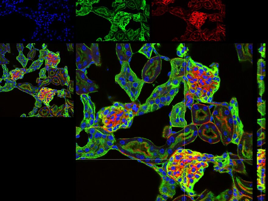
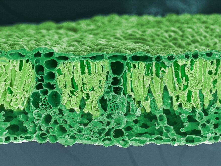
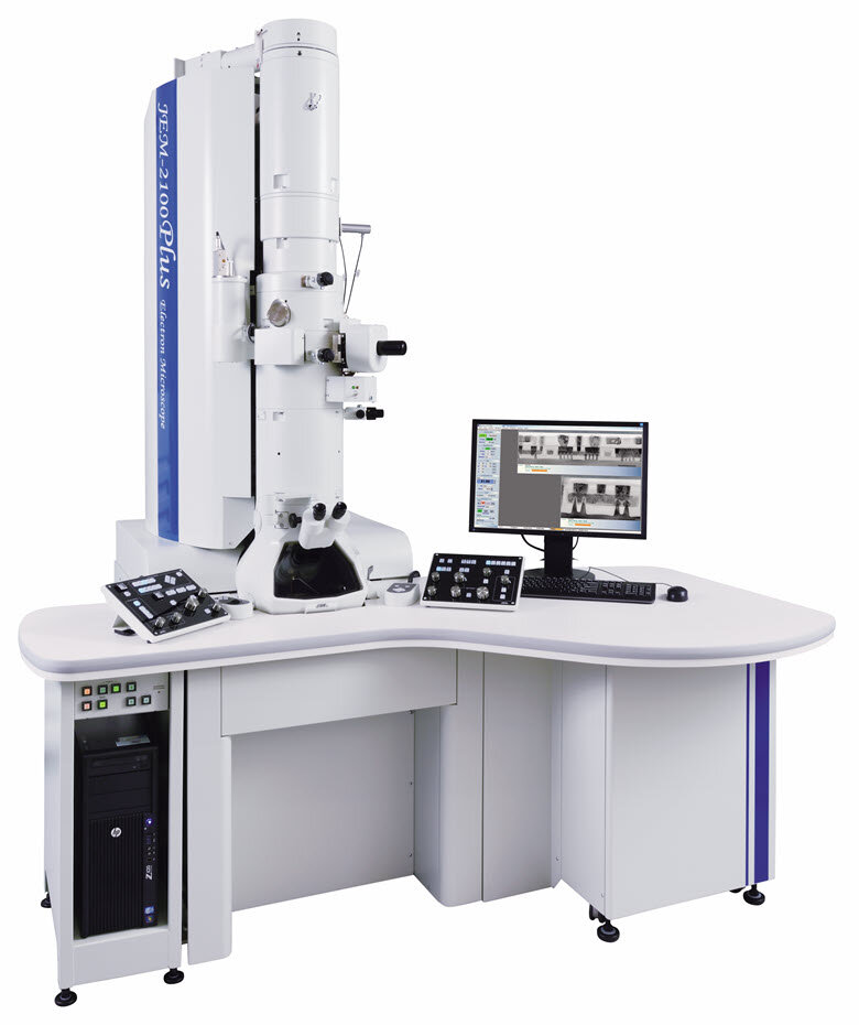
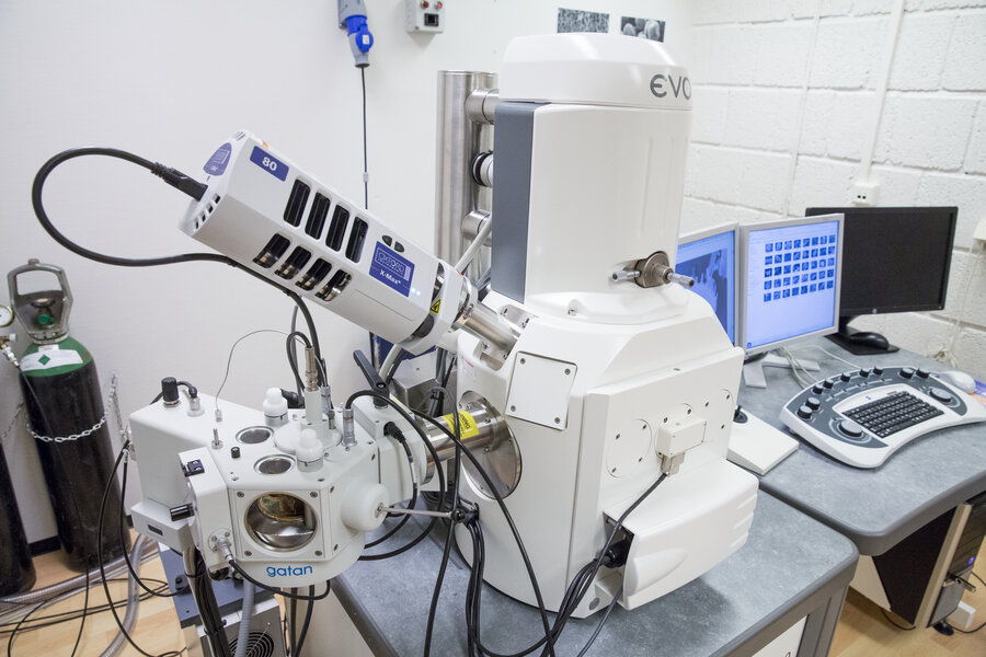
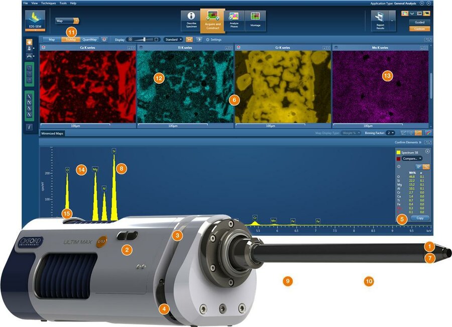
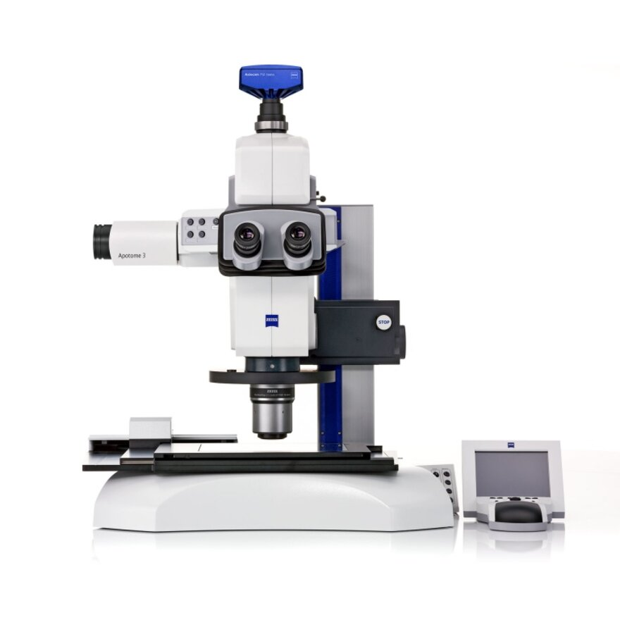
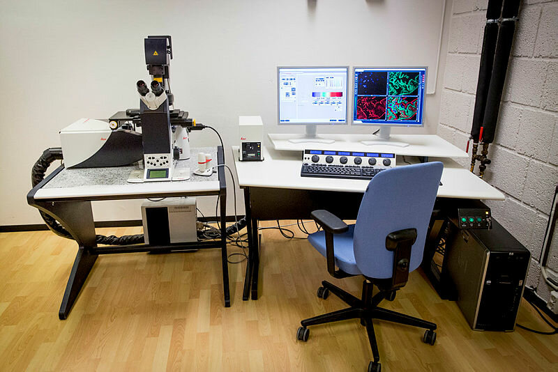
Training and technical support in a variety of microscopy techniques, including light, confocal, scanning electron (SEM) and high-resolution scanning transmission electron microscopy (TEM).
The Core Facility offers a comprehensive service, including imaging and custom sample preparation.
We can provide guidance and technical expertise on the use of our entire infrastructure, to provide a low threshold for all users whether experienced or not.
The Imaging Centre is accessible to all internal and external users, both academic and industrial.
The Imaging Centre has moved the Veterinary Building, May 2025.
Contact information:
E-mail: imaging.centre@nmbu.no
Phone: 67 23 27 61/62, 92 41 72 43 or 48 25 98 43
Visiting address:
Elizabeth Stephansens vei 15,
Hippocampus,
The Veterinary Building
Postal address:
Norwegian University of Life Sciences
P.O. Box 5003
NO-1432 Ås
Norway
Opening hours:
Kl. 08:00 to 15.45
15. May to 14. September:
kl. 08:00 to 15.00
News
Booking system and prices
Courses
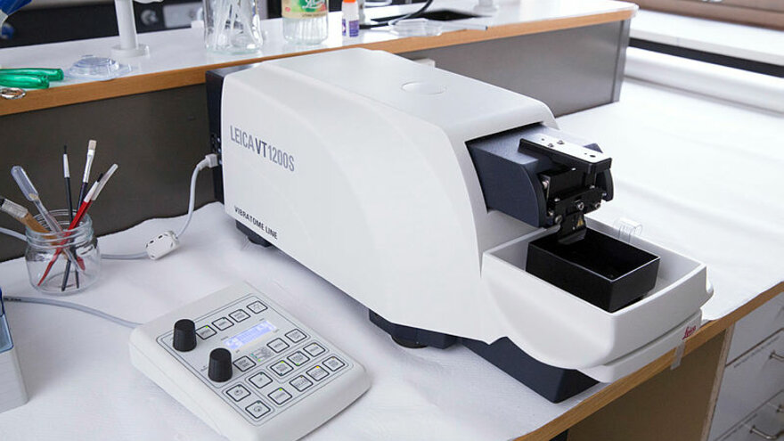
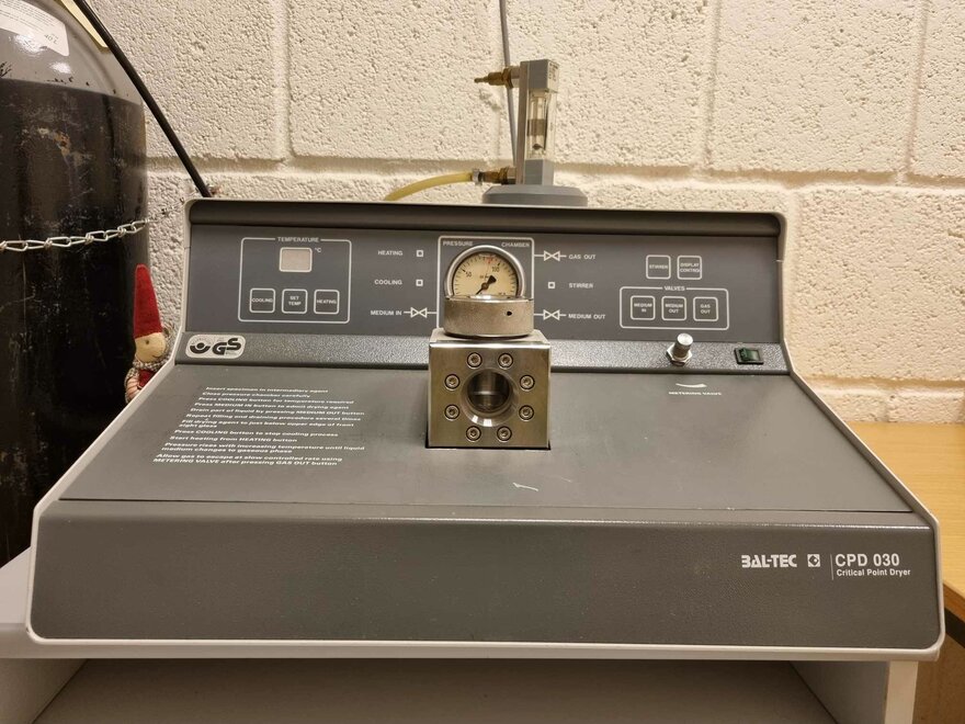
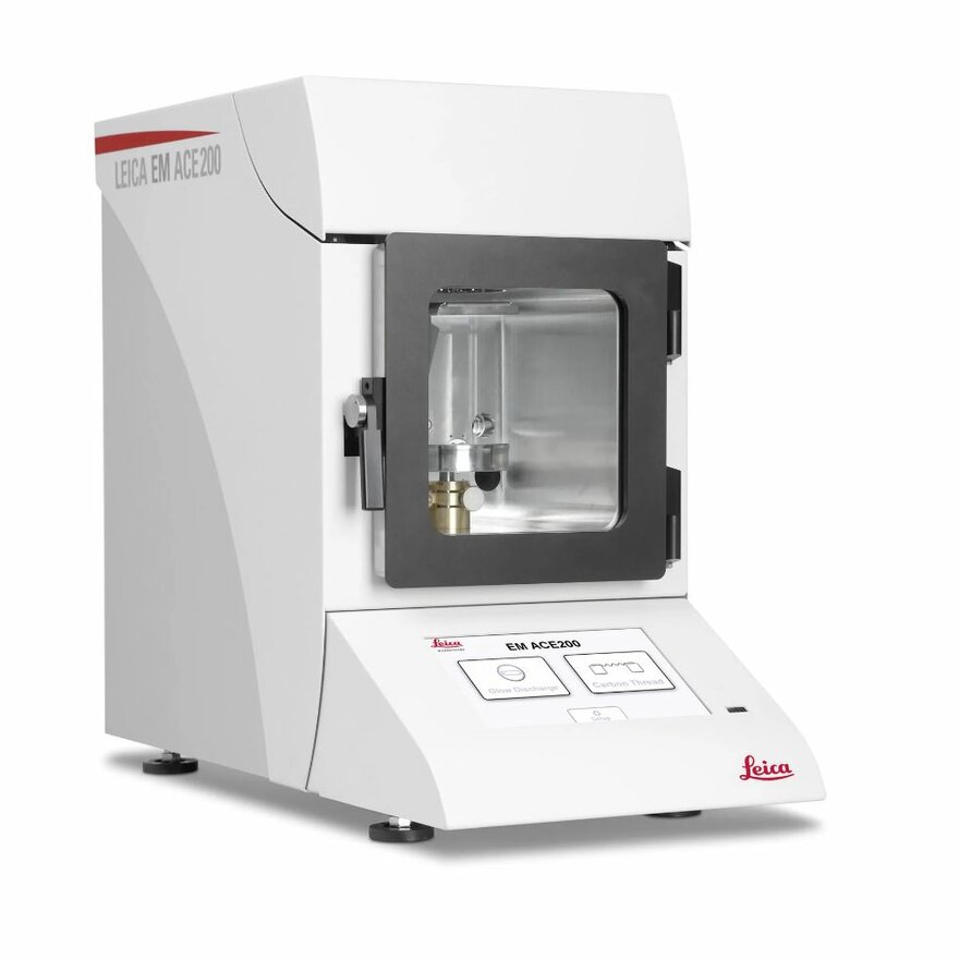
Equipment


Employees at the Imaging Centre